Diagram Of Hip Muscles And Ligaments / Pin on Diagram. This causes them to become tighter when the joint is extended. Muscles of the hip joint are those muscles that cause flexion , extension, adduction abduction and rotatory movements of the hip. Most modern anatomists define 17 of these muscles, although some additional muscles may sometimes be considered. Knee assessment and hip mechanics learn how hip and pelvis. The inguinal ligament supports the muscles that run inferior to its fibers, including the iliopsoas and pectineus muscles of the hip.
Iliac crest and iliolumbar ligament. Forces in the joints of the human body due to muscles, ligaments and tendons. Even if you're not an athlete, the state of your hip ligaments, tendons, and muscles play an important role in the function of the hip. Iliofemoral ligament is the most formidable ligament of body and prevents the trunk from falling backwards in the standing position. Diagram of hip muscles and ligaments.
Hip Anatomy | eOrthopod.com from eorthopod.com Flexes hip and/or lumbar spine. • justify the actions of the hip muscles through knowledge of the muscle's proximal and distal attachments. Each hip bone consists of the ilium, ischium, and pubic bone. The external surface of the capsule of the hip is reinforced by a set of thick and important ligaments: Want to read the whole page? Iliofemoral ligament is the most formidable ligament of body and prevents the trunk from falling backwards in the standing position. All three strengthen the joint by preventing excessive range of movement and also become taut when the joint is extended in order to stabilize it. Magnetic resonance musculoskeletal models are helpful in understanding the reduction of the.
A strong fibrous capsule encloses the hip joint that aids in the maintenance of hip stability.
This causes them to become tighter when the joint is extended. Hip bony bits, pelvis and ligaments. Synovial joints are often supported and reinforced by surrounding ligaments, which limit movement to prevent injury. Ligaments of the hip elma. The pelvis connects the lower extremity to the trunk, protects abdominal and pelvic organs, and provides attachment to muscles the pelvic joints and the organs are supported by muscles and ligaments (including the urogenital diaphragm). A strong fibrous capsule encloses the hip joint that aids in the maintenance of hip stability. The iliofemoral ligament, the pubofemoral ligament and the ischiofemoral ligament all act to strengthen the mobile capsule. Muscles creating the movements of the hip joint. As previously stated, the muscles of the hip joint can contribute to movement in several different (3). The psoas major unites with the iliacus at the level of the inguinal ligament and crosses the hip joint to insert on the lesser trochanter of the femur. Approaching the muscular anatomy around the hip can be undertaken in a number of ways. Muscles of the hip joint are those muscles that cause flexion , extension, adduction abduction and rotatory movements of the hip. Iliofemoral ligament is the most formidable ligament of body and prevents the trunk from falling backwards in the standing position.
Several muscles cross the front of the hip and create hip flexion, pulling the thigh and trunk toward each other, but probably the most important is the when the cast is removed after six or eight weeks, the soft tissues around the elbow (muscles, tendons, ligaments, and even skin) will have shortened. Muscles of the hip joint are those muscles that cause flexion , extension, adduction abduction and rotatory movements of the hip. Gluteus maximus, gluteus medius, gluteus minimus, tensor fasciae latae inner hip. Ligaments and tendons are fibrous bands of connective tissue that attach to bone. As previously stated, the muscles of the hip joint can contribute to movement in several different (3).
Ultimate hip joint anatomy in one picture! | Hip anatomy, Joints anatomy, Hip joint anatomy from i.pinimg.com This causes them to become tighter when the joint is extended. The inguinal ligament supports the muscles that run inferior to its fibers, including the iliopsoas and pectineus muscles of the hip. Together with the infrahyoid muscles (discussed below) these muscles fix the hyoid bone and this enables the hyoid bone to serve as a depress ribs. The inguinal ligament is often incorrectly referred to as a fallopian ligament or poupart's ligament. While walking, where k is the body weight. Ligaments aid in joint stability during rest and movement and help prevent injury from hyperextension and hyperflexion (excessive movements). Magnetic resonance musculoskeletal models are helpful in understanding the reduction of the. Ligaments are soft tissue structures that connect bones to bones.
The psoas major unites with the iliacus at the level of the inguinal ligament and crosses the hip joint to insert on the lesser trochanter of the femur.
Flexes hip and/or lumbar spine. That study illustrated the complex interaction between native hip structures in the context of hip arthroplasty (i.e., capsule, ligaments, and muscles) and joint mechanics specific to a surgical. A small opening in the muscles and connective tissues of the abdomen — known as the superficial inguinal ring — is located just superior to the inguinal ligament. • justify the actions of the hip muscles through knowledge of the muscle's proximal and distal attachments. In addition, the muscles and ligaments. Approaching the muscular anatomy around the hip can be undertaken in a number of ways. All three strengthen the joint by preventing excessive range of movement and also become taut when the joint is extended in order to stabilize it. As previously stated, the muscles of the hip joint can contribute to movement in several different (3). Together with the infrahyoid muscles (discussed below) these muscles fix the hyoid bone and this enables the hyoid bone to serve as a depress ribs. Ligaments and tendons are fibrous bands of connective tissue that attach to bone. Knee assessment and hip mechanics learn how hip and pelvis. Tibialis anterior, extensor hallicus longus, fibularis tertius, and extensor digitorum longus and extensor digitorum brevis muscle; The external surface of the capsule of the hip is reinforced by a set of thick and important ligaments:
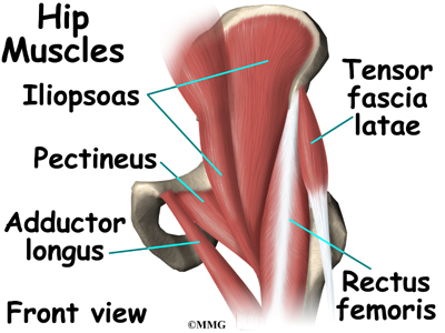
The acetabulum is a concave area in the pelvis, into which the femoral head fits. Knee assessment and hip mechanics learn how hip and pelvis. Synovial joints are often supported and reinforced by surrounding ligaments, which limit movement to prevent injury. Hip bony bits, pelvis and ligaments. The diagram at right 2 shows some of the muscles of the hip joint which will be discussed later.
Bones, Ligaments & Joints - Atlas of Anatomy | Hip bones, Leg bones, Anatomy from i.pinimg.com As noted above, the stability of the hip joint is directly related to its muscles and ligaments. Even if you're not an athlete, the state of your hip ligaments, tendons, and muscles play an important role in the function of the hip. A small opening in the muscles and connective tissues of the abdomen — known as the superficial inguinal ring — is located just superior to the inguinal ligament. Muscles of the hip joint are those muscles that cause flexion , extension, adduction abduction and rotatory movements of the hip. Related online courses on physioplus. In human anatomy, the muscles of the hip joint are those muscles that cause movement in the hip. Gluteus maximus, gluteus medius, gluteus minimus, tensor fasciae latae inner hip. Learn vocabulary, terms and more with flashcards, games and other study tools.
The external surface of the capsule of the hip is reinforced by a set of thick and important ligaments:
Tags ischial tuberosity, sacrotuberous ligament. Learn vocabulary, terms and more with flashcards, games and other study tools. Muscles surrounding the joint influence its stability and motion. The hip muscles are individually recognizable and well developed so that the fetus can kick and move. Ligaments and tendons are fibrous bands of connective tissue that attach to bone. Ligaments are fibrous bands or sheets of connective tissue linking two or more bones, cartilages, or structures together. Receives the greatest stresses of the ligaments. The muscles involved in hip motion are attached to the joint at these trochanters. Related online courses on physioplus. Magnetic resonance musculoskeletal models are helpful in understanding the reduction of the. Even if you're not an athlete, the state of your hip ligaments, tendons, and muscles play an important role in the function of the hip. • justify the actions of the hip muscles through knowledge of the muscle's proximal and distal attachments. The inguinal ligament is often incorrectly referred to as a fallopian ligament or poupart's ligament.
The iliofemoral ligament, the pubofemoral ligament and the ischiofemoral ligament all act to strengthen the mobile capsule hip muscles diagram. Learn vocabulary, terms and more with flashcards, games and other study tools.
Share :
Post a Comment
for "Diagram Of Hip Muscles And Ligaments / Pin on Diagram"


Post a Comment for "Diagram Of Hip Muscles And Ligaments / Pin on Diagram"